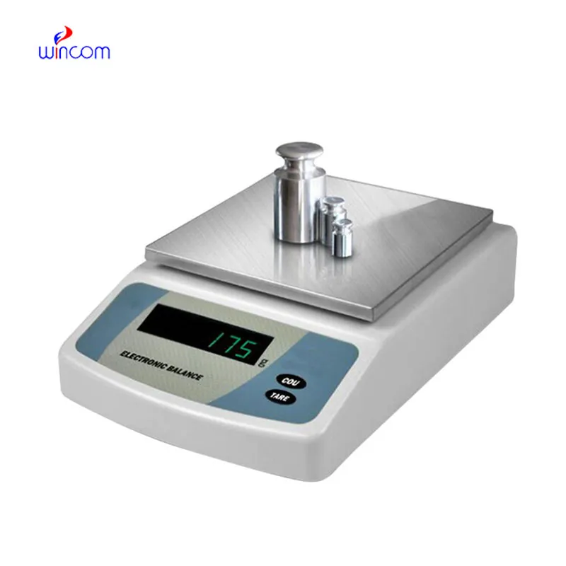
The use of analytic balance in pharmaceutical laboratories is crucial for the accurate measurement of both active substances and excipients. Its extraordinary accuracy eliminates the possibility of formulation errors and makes regulatory compliance easier. analytic balance are employed by laboratory staff for daily quality control, validation of batches, and research activities. Adding analytic balance to the laboratory operations not only the consistency but also the reproducibility and the accuracy of the results for clinical trials and research applications are assured.

In microbiology labs, analytic balance is utilized for the preparation of culture media and analytical additives. Accurate weighing guarantees that the nutrient composition for microbial growth and testing is consistent. This application helps to produce reliable cultures, conduct antimicrobial studies, and do infection research. By keeping the mass control precise, analytic balance supports reproducibility in microbiological workflows in clinical laboratories.
In forthcoming times, analytic balance is foreseen to assimilate additional digital interrelation among hospital laboratories. Improved information interchange will make it possible to transfer weighing records directly to laboratory information systems, thus cutting down paper work done manually. This advancement will contribute to traceability, audit readiness, and efficiency in clinical workflows. When hospitals keep on embracing intelligent laboratory infrastructure, analytic balance will be an essential element of connected analytical ecosystems, helping with real-time monitoring and centralized data management.
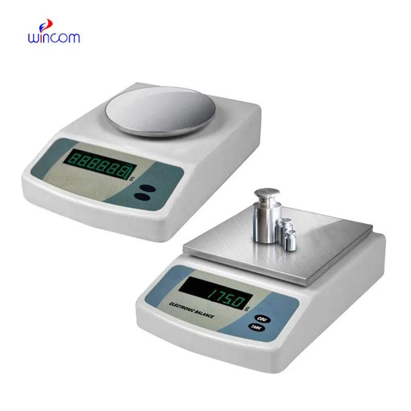
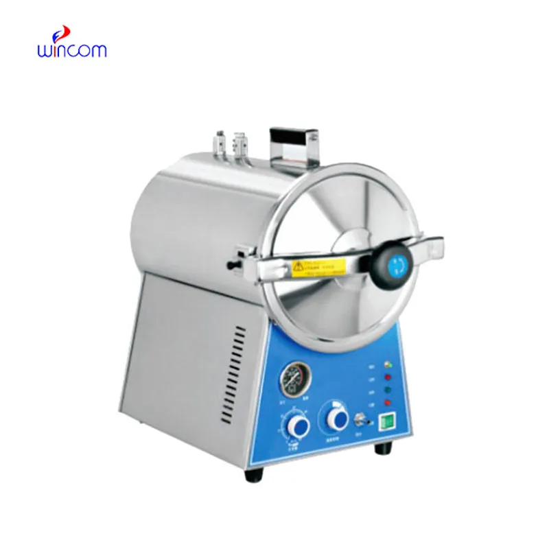
In order to keep analytic balance in a good condition consistent calibration practices are needed that follow hospital laboratory protocols. Scheduled calibration checks are performed to maintain the reliability of measurements during daily activities involving analysis. Conditions in the environment such as temperature and the amount of air that moves around should be kept under control so as to prevent drift. The people operating the machines should make sure that there are no sudden changes in load and that the weighing pan is not subjected to excessive force. Through adhering to controlled handling practices, analytic balance is always trusted for pharmaceutical preparation and medical research activities.
Balance is crucial in the various ranges of hospital and clinical laboratories for the preparation of patient samples to be analyzed. Because weighing correctly provides proper reagent ratios, it ensures consistent dilutions and valid diagnostic test results. Laboratory staff can achieve a huge array of quality standards in sample preparation with analytic balance, being assured of reliable clinical diagnostics, treatment monitoring, and patient safety by means of precise measurement of laboratory materials.
Q: What is the impact of temperature on the performance of analytical balance? A: The changes in temperature can lead to drift and weighing inconsistency. Q: Are analytical balances the only ones used in research laboratories? A: They are very important also for other processes such as sample preparation and improving the accuracy of the experiment. Q: How long does it usually take for an analytical balance to warm up? A: Warm-up times differ from one model to another, but an adequate stabilizing period increases the reliability of the measurement. Q: Is it possible for analytical balances to save weighing data? A: Internal memory or external data transfer are the two ways in which many models can achieve this feature. Q: Would it be necessary to undergo training if one wants to operate an analytical balance? A: Basic laboratory training will be enough to make sure that the balance is being used correctly.
We’ve been using this mri machine for several months, and the image clarity is excellent. It’s reliable and easy for our team to operate.
The water bath performs consistently and maintains a stable temperature even during long experiments. It’s reliable and easy to operate.
To protect the privacy of our buyers, only public service email domains like Gmail, Yahoo, and MSN will be displayed. Additionally, only a limited portion of the inquiry content will be shown.
Could you please provide more information about your microscope range? I’d like to know the magnif...
I’d like to inquire about your x-ray machine models. Could you provide the technical datasheet, wa...
E-mail: [email protected]
Tel: +86-731-84176622
+86-731-84136655
Address: Rm.1507,Xinsancheng Plaza. No.58, Renmin Road(E),Changsha,Hunan,China

 af
af
 es
es
 ar
ar
 tr
tr
 sw
sw
 pt
pt
 th
th
 ur
ur
 bn
bn
 ne
ne
 vi
vi
 km
km
 lo
lo
 de
de
 ru
ru
 fi
fi
 nl
nl
 fa
fa
 fr
fr
 ko
ko