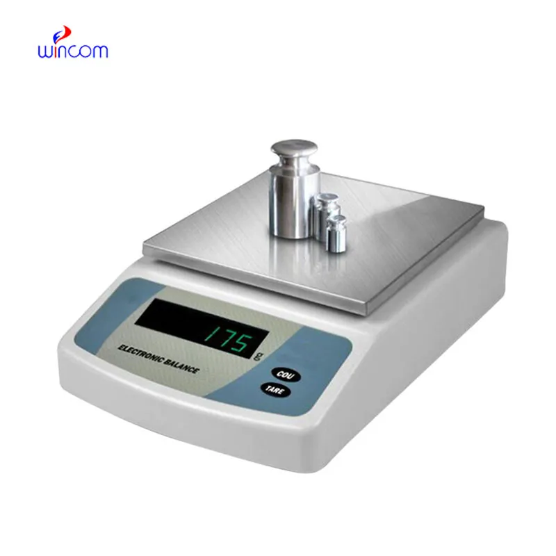
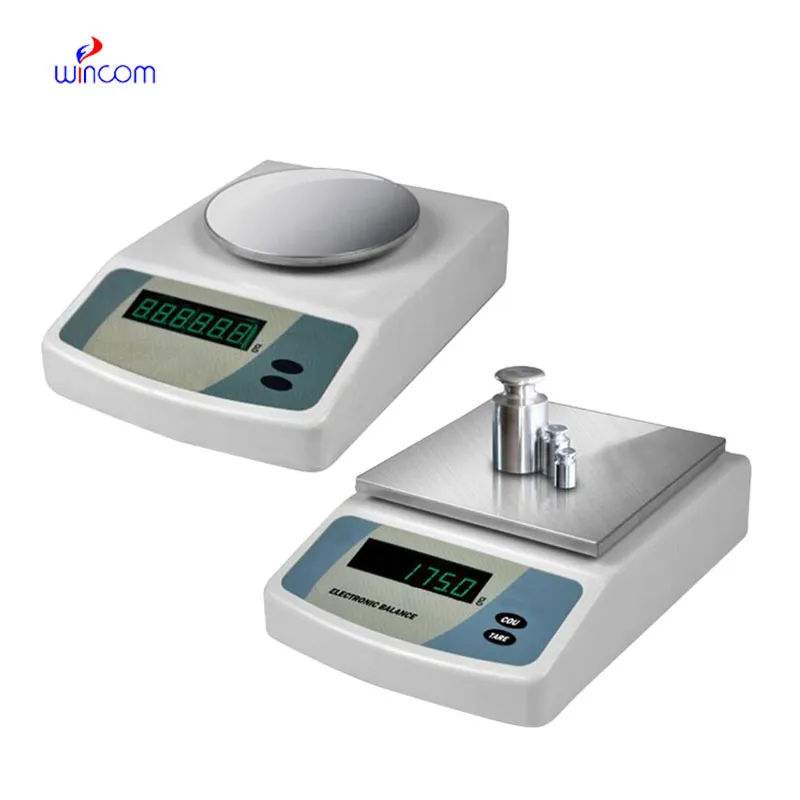
analytical balance for laboratory ensures accurate weighing of clinical sample preparation, research studies, and hospital and laboratory medication formulation. It has an application envisaged to measure even the tiniest amounts with excellent sensitivity and repeatability. analytical balance for laboratory is the instrument laboratory technicians trust for maintaining the highest level of accuracy in analysis, validation of experimental practices, and patients' care support. The use of this instrument in the lab operations not only guarantees results that are reliable but also creates a consistent workflow and quality control which is improved in both the diagnostic and research environments.

analytical balance for laboratory is commonly used in the compounding of medicinal and chemical substances in small quantities in hospital pharmacy laboratories. Mass control with high precision is critical when dealing with active pharmaceutical ingredients in micro or milligram quantities. This application helps to prepare exact doses, internal validation, and experimental drug research. By allowing for repeatable measurements, analytical balance for laboratory helps pharmacists and researchers in controlling formulation processes, which makes it easier to get consistency and reliability throughout hospital medication development workflows.
The future of analytical balance for laboratory in medical labs will put more focus on environmental stability. The advanced vibration suppression and temperature control capabilities will support precise operation even in the most crowded hospital areas. This change will make it possible to locate analytical balance for laboratory nearer to the clinical workstations and this, in turn, will result in a reduction of sample transport time. Rather than moving to simpler hospital environments, analytical balance for laboratory will continue to provide quick analytical preparation support and will also maintain high measurement consistency.
In order to keep analytical balance for laboratory in a good condition consistent calibration practices are needed that follow hospital laboratory protocols. Scheduled calibration checks are performed to maintain the reliability of measurements during daily activities involving analysis. Conditions in the environment such as temperature and the amount of air that moves around should be kept under control so as to prevent drift. The people operating the machines should make sure that there are no sudden changes in load and that the weighing pan is not subjected to excessive force. Through adhering to controlled handling practices, analytical balance for laboratory is always trusted for pharmaceutical preparation and medical research activities.
The precision of analytical balance for laboratory is achieved only in a very controlled environment, which implies regulation of temperature, humidity, and vibration to a minimum level. These parameters are continuously monitored by laboratory technicians to avoid any errors in measurements. analytical balance for laboratory technique provides highly accurate weighing of tiny samples in severe conditions, thus supporting laboratory experiments and hospital-grade analyses of sensitive tests or research that demands careful sample handling.
Q: What is the main purpose of an Analytical Balance? A: Its purpose is mainly to measure very tiny sample masses with the utmost precision in laboratories and hospitals. Q: What is the typical weighing range of an Analytical Balance? A: The weighing range for the majority of analytical balances is from 0 up to some grams with a resolution of micrograms or milligrams. Q: What environmental controls are necessary for an Analytical Balance's operation? A: Airflow, vibration, and temperature changes should not only be avoided but also prevented in the room where the scale is situated. Q: Is an Analytical Balance permitted in a hospital laboratory? A: Yes, it has indeed found widespread usage for the preparation of reagents, calibra¬tion, and drug development applications. Q: What should be the frequency of calibration for an Analytical Balance? A: The calibration interval is subject to the degree of use and the particular laboratory requirements.
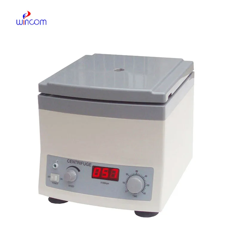
The centrifuge operates quietly and efficiently. It’s compact but surprisingly powerful, making it perfect for daily lab use.
The water bath performs consistently and maintains a stable temperature even during long experiments. It’s reliable and easy to operate.
To protect the privacy of our buyers, only public service email domains like Gmail, Yahoo, and MSN will be displayed. Additionally, only a limited portion of the inquiry content will be shown.
Could you please provide more information about your microscope range? I’d like to know the magnif...
Could you share the specifications and price for your hospital bed models? We’re looking for adjus...
E-mail: [email protected]
Tel: +86-731-84176622
+86-731-84136655
Address: Rm.1507,Xinsancheng Plaza. No.58, Renmin Road(E),Changsha,Hunan,China

 af
af
 es
es
 ar
ar
 tr
tr
 sw
sw
 pt
pt
 th
th
 ur
ur
 bn
bn
 ne
ne
 vi
vi
 km
km
 lo
lo
 de
de
 ru
ru
 fi
fi
 nl
nl
 fa
fa
 fr
fr
 ko
ko