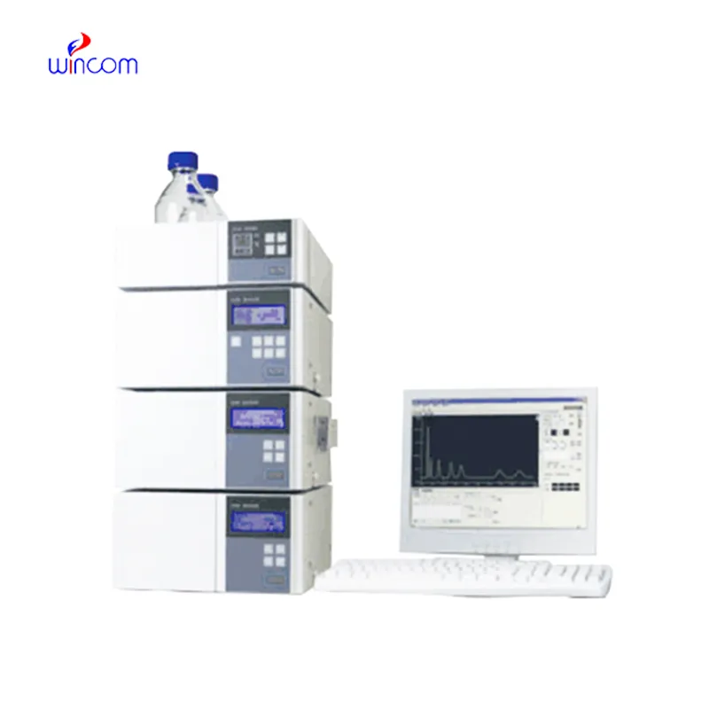
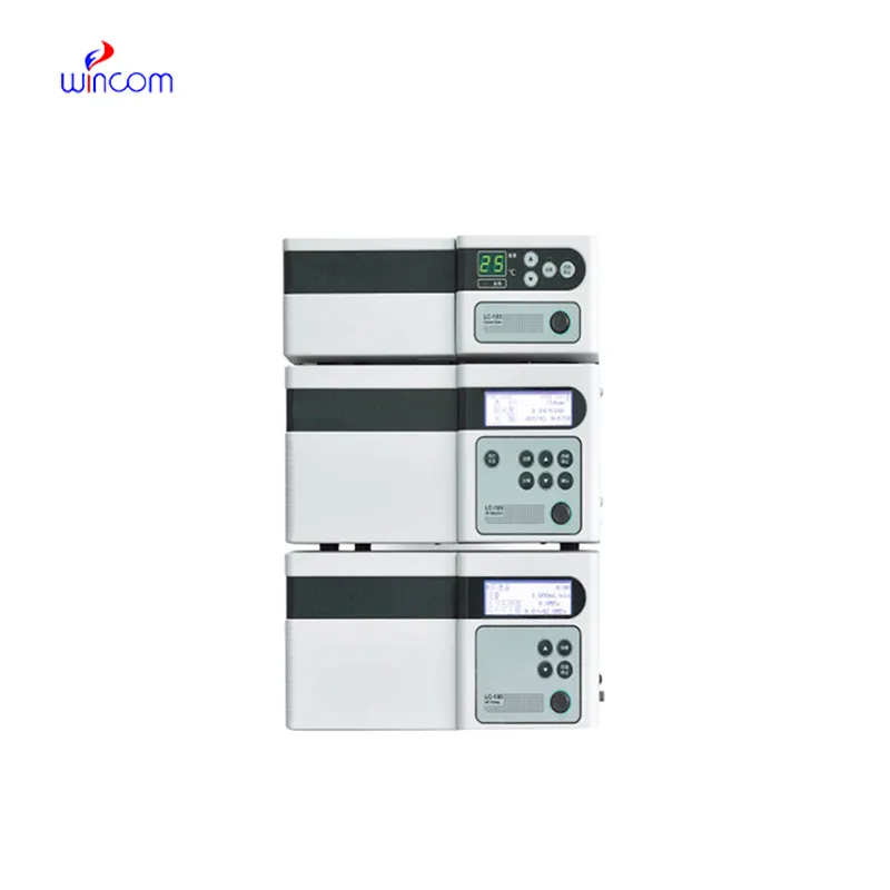
Hospitals and biomed research centers employ automated liquid chromatography that help optimize patient testing and lab work. By being able to distinguish, measure, and analyze drugs, metabolites, and biomolecules, automated liquid chromatography is a necessary tool in patient testing. Lab professionals incorporate automated liquid chromatography into lab work on a daily basis. Reproducibility and analytical ability make automated liquid chromatography an irreplaceable tool in assisting with patient testing.

automated liquid chromatography allows the personnel of hospitals and laboratories to keep an eye on the presence of environmental pollutants in sterile drugs. It purifies and recognizes the remaining solvents, preservatives, and other possible impurities thus, confirming safety and meeting the requirements of regulatory authorities. This technology is vital in the battle against exposing patients to toxic agents.
The future of automated liquid chromatography stresses the integration of hospital information systems and electronic medical records. The analysis of patient samples will be automatically included in the clinical workflows. Increased automation, AI-based interpretation, and better sensitivity will put automated liquid chromatography at the center of the laboratory operations and patient care that is focused on the patient's needs.
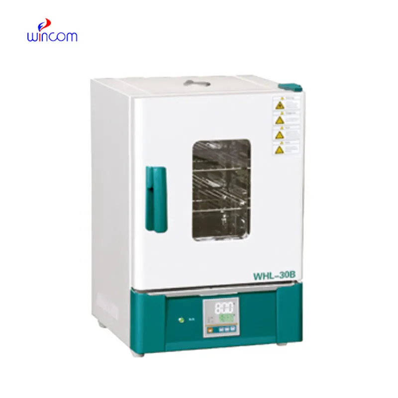
Preventive maintenance is automated liquid chromatography that play a very important role in clinical and hospital laboratories. The routine performance of flushing columns, cleaning injector valves, and monitoring pressure stability extends the life of the system. The laboratory staff is required to keep records of maintenance activities, replace consumables in a timely manner, and use solvents that are compatible. All of these practices are essential for the instruments' performance retention, lifespan extension, and high-quality analytical results, both in patient sample testing and research.
The automated liquid chromatography is the backbone of quality control and drug analysis in the pharmaceutical sector. It was able to identify the active ingredients and side products in a very complex, but at the same time, accurate manner. With the choice of proper columns and mobile phases, specialists can isolate the components in both a very efficient and a very constant manner. automated liquid chromatography data is very often requested by regulatory bodies in order to confirm quality of the batch and keep the patients safe. Its accuracy is the mainstay for dosage checking and stability studies. The capability of detecting substances at the trace level renders automated liquid chromatography as the most used and sometimes the only method in drug development, production supervision, and formulation research, thus compliance with industry standards being ensured.
Q: Do you need special training for HPLC operation? A: The answer is yes, training is a prerequisite to accurately and safely using pumps, columns, and detectors. Q: What type of maintenance does HPLC have? A: It requires cleaning, flushing, and inspection of all components as well as calibrating. Q: Is it possible to use HPLC in drug monitoring? A: Sure, it is a common practice in hospitals to monitor the levels of therapeutic drugs and also to identify metabolites in the samples taken from the patients. Q: What is the duration of analysis using HPLC in a typical case? A: The analysis time can range from a few minutes to more than an hour depending on the nature of the sample and the kind of column used. Q: Is HPLC a good choice for environmental testing? A: Yes, it can be used to find out the presence of pollutants, pesticides, and other harmful substances in water, soil, and air samples.
The delivery bed is well-designed and reliable. Our staff finds it simple to operate, and patients feel comfortable using it.
The microscope delivers incredibly sharp images and precise focusing. It’s perfect for both professional lab work and educational use.
To protect the privacy of our buyers, only public service email domains like Gmail, Yahoo, and MSN will be displayed. Additionally, only a limited portion of the inquiry content will be shown.
We’re currently sourcing an ultrasound scanner for hospital use. Please send product specification...
We are planning to upgrade our imaging department and would like more information on your mri machin...
E-mail: [email protected]
Tel: +86-731-84176622
+86-731-84136655
Address: Rm.1507,Xinsancheng Plaza. No.58, Renmin Road(E),Changsha,Hunan,China

 af
af
 es
es
 ar
ar
 tr
tr
 sw
sw
 pt
pt
 th
th
 ur
ur
 bn
bn
 ne
ne
 vi
vi
 km
km
 lo
lo
 de
de
 ru
ru
 fi
fi
 nl
nl
 fa
fa
 fr
fr
 ko
ko