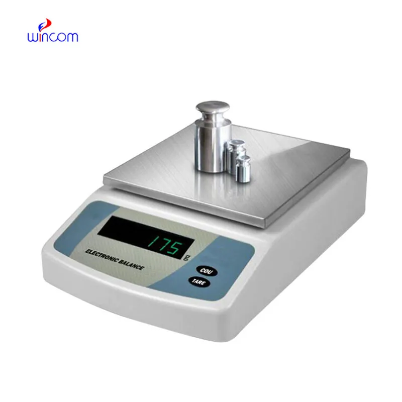
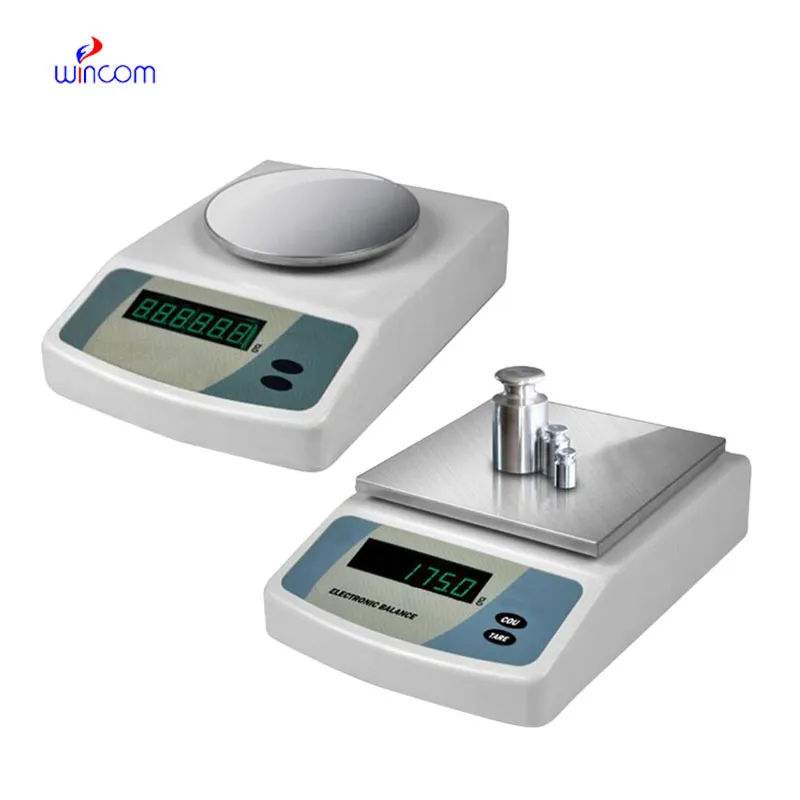
electronic scale balance ensures accurate weighing of clinical sample preparation, research studies, and hospital and laboratory medication formulation. It has an application envisaged to measure even the tiniest amounts with excellent sensitivity and repeatability. electronic scale balance is the instrument laboratory technicians trust for maintaining the highest level of accuracy in analysis, validation of experimental practices, and patients' care support. The use of this instrument in the lab operations not only guarantees results that are reliable but also creates a consistent workflow and quality control which is improved in both the diagnostic and research environments.

Hospitals' analytical laboratories use electronic scale balance during the production of internal standards for instrument testing methods. Very accurate mass input is a must to maintain uniformity over different analytical runs. This use case allows for comparison of still data, traceability, and monitoring of analytical performance over a long period. By allowing exact preparation of reference materials, electronic scale balance boosts measurement confidence in all phases of hospital laboratory work.
The future application of electronic scale balance will be broadened in education laboratories at teaching hospitals. The training provided to lab techs and medical researchers will be accomplished with the help of advanced simulation modes and guided measurement functions. This revolution will offer the medical student the chance to learn practically while the doctor will continue to rely on the precision of the instruments in the lab.
electronic scale balance in clinics is, however, maintained through regular performance verification conducted in the laboratories. Certified test weights are used to prove the reliability of the measurements with the passage of time. Verification activities being documented also implies good traceability and makes it easier for internal audits to take place. By making verification part of the routine maintenance, hospitals make sure that electronic scale balance still gives trustworthy results for research and diagnostic workflows.
Medical research laboratories rely on electronic scale balance to determine the weight of samples for their experimental procedures. No matter if weighing chemicals, biomolecules, or powders, accuracy is vital for repeatability. Scientific workers apply electronic scale balance so that slight changes in sample weight do not affect the validity of the results. The application of this tool helps to enhance the reputation of the laboratory, the quality of the experiments, and the uniformity of the research conducted in hospitals or drug companies.
Q: What is the main purpose of an Analytical Balance? A: Its purpose is mainly to measure very tiny sample masses with the utmost precision in laboratories and hospitals. Q: What is the typical weighing range of an Analytical Balance? A: The weighing range for the majority of analytical balances is from 0 up to some grams with a resolution of micrograms or milligrams. Q: What environmental controls are necessary for an Analytical Balance's operation? A: Airflow, vibration, and temperature changes should not only be avoided but also prevented in the room where the scale is situated. Q: Is an Analytical Balance permitted in a hospital laboratory? A: Yes, it has indeed found widespread usage for the preparation of reagents, calibra¬tion, and drug development applications. Q: What should be the frequency of calibration for an Analytical Balance? A: The calibration interval is subject to the degree of use and the particular laboratory requirements.
The centrifuge operates quietly and efficiently. It’s compact but surprisingly powerful, making it perfect for daily lab use.
The hospital bed is well-designed and very practical. Patients find it comfortable, and nurses appreciate how simple it is to operate.
To protect the privacy of our buyers, only public service email domains like Gmail, Yahoo, and MSN will be displayed. Additionally, only a limited portion of the inquiry content will be shown.
I’d like to inquire about your x-ray machine models. Could you provide the technical datasheet, wa...
I’m looking to purchase several microscopes for a research lab. Please let me know the price list ...
E-mail: [email protected]
Tel: +86-731-84176622
+86-731-84136655
Address: Rm.1507,Xinsancheng Plaza. No.58, Renmin Road(E),Changsha,Hunan,China

 af
af
 es
es
 ar
ar
 tr
tr
 sw
sw
 pt
pt
 th
th
 ur
ur
 bn
bn
 ne
ne
 vi
vi
 km
km
 lo
lo
 de
de
 ru
ru
 fi
fi
 nl
nl
 fa
fa
 fr
fr
 ko
ko