The fetal heartbeat doppler nearby, being designed for flexibility, brings the user a variety of imaging modes such as B-mode, M-mode, and color Doppler to use. Because of its small size, it can be easily moved from one place to another and that is why it can be used for examining patients in bed. The fetal heartbeat doppler nearby provides good imaging quality, which can be depended upon in cases of routine diagnostics, fieldwork, and emergency medical interventions.

The fetal heartbeat doppler nearby is a very significant diagnosis tool used in obstetrics for fetal monitoring and pregnancy development detection. It indirectly affects the cardiology field by providing information about the hearts and blood flow dynamics. Furthermore, the fetal heartbeat doppler nearby is very important in diagnosing abdominal problems, especially issues with the liver, kidneys, and gallbladder. Still, it is also being used in musculoskeletal diagnoses for spotting ligament and tendon injuries.

The coming years will see the evolution of the fetal heartbeat doppler nearby into an independent and adaptable imaging solution. The increased level of automation will eliminate the need for human input. The fetal heartbeat doppler nearby may include predictive model components that will help healthcare providers to identify probable risks to an individual's health.

In order to retain the accuracy of the fetal heartbeat doppler nearby, it is important for operators to check the cables and connections of the transducers for evidence of wear. After each use, the surfaces should be wiped clean using non-abrasive cleaners. The fetal heartbeat doppler nearby should be turned off properly and covered to prevent dust from collecting. Regular checks by trained personnel should be done.
With the advanced imaging technology, the fetal heartbeat doppler nearby provides physicians unobstructed and precise images of internal organs. It is employed in the early disease diagnosis as well as in patient tracking. The fetal heartbeat doppler nearby functions by utilizing sound wave reflections to generate dynamic images, qualifying it as an essential tool in modern medical diagnostics. Through the fetal heartbeat doppler nearby, fast, non-invasive testing is facilitated for real-time assessment to support clinical decisions.
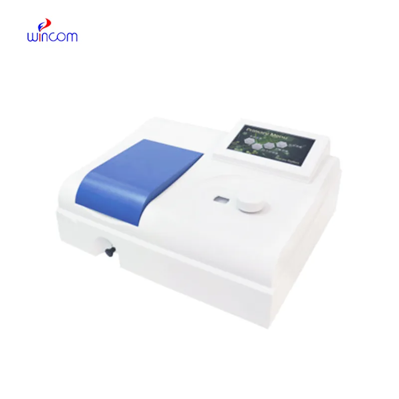
Q: What are the main maintenance requirements for the ultrasound scannert? A: Regular cleaning, proper probe handling, and scheduled inspections help maintain optimal performance. Q: How often should the ultrasound scannert be calibrated? A: Calibration frequency depends on usage levels, but periodic professional checks are recommended. Q: Is the ultrasound scannert suitable for pediatric use? A: Yes, it provides gentle, non-invasive imaging ideal for neonatal and pediatric diagnostics. Q: Does the ultrasound scannert support wireless connectivity? A: Many models include Wi-Fi or Bluetooth features for data sharing and device integration. Q: What materials are used in the ultrasound scannert construction? A: It is built with durable medical-grade components designed to withstand continuous clinical use.
The hospital bed is well-designed and very practical. Patients find it comfortable, and nurses appreciate how simple it is to operate.
This x-ray machine is reliable and easy to operate. Our technicians appreciate how quickly it processes scans, saving valuable time during busy patient hours.
To protect the privacy of our buyers, only public service email domains like Gmail, Yahoo, and MSN will be displayed. Additionally, only a limited portion of the inquiry content will be shown.
We’re interested in your delivery bed for our maternity department. Please send detailed specifica...
I’d like to inquire about your x-ray machine models. Could you provide the technical datasheet, wa...
E-mail: [email protected]
Tel: +86-731-84176622
+86-731-84136655
Address: Rm.1507,Xinsancheng Plaza. No.58, Renmin Road(E),Changsha,Hunan,China

 af
af
 es
es
 ar
ar
 tr
tr
 sw
sw
 pt
pt
 th
th
 ur
ur
 bn
bn
 ne
ne
 vi
vi
 km
km
 lo
lo
 de
de
 ru
ru
 fi
fi
 nl
nl
 fa
fa
 fr
fr
 ko
ko