With video displayed through beamforming and noise-filtering technology, the handheld ultrasound scanner is able to present an image that is very sharp and stable. Easy-to-use touch-screen controls help to streamline the process while rapid image rendering is guaranteed by the fast processing. Equipped for contemporary healthcare settings, the handheld ultrasound scanner is capable of working with both 2D and Doppler imaging.

The handheld ultrasound scanner is a tool that medical professionals are using in different departments like pediatrics for the measurement of the size of the organs and neurology for appreciating the anatomy of the soft tissues. It makes it easy for anesthesiologists to perform nerve blocks and vascular access procedures. The handheld ultrasound scanner increase the speed and the trust of the diagnosis during both routine checkups and highly technical interventions.

The future of the handheld ultrasound scanner may include enhancements based on artificial intelligence algorithms that can improve images and offer automatic measurements. Better transducer technologies may offer higher resolution and increased sensitivity. The handheld ultrasound scanner may have an increasing role to play in personalized medicine because it may offer continuous monitoring capabilities.

The daily upkeep of the handheld ultrasound scanner involves cleaning, inspection, and proper storage. The removal of the gel residue from the probes should be accomplished as soon as the analysis has been carried out. The cooling vents of the device should always be unblocked. The handheld ultrasound scanner needs annual professional servicing in order to remain accurate.
Built for performance and accuracy, the handheld ultrasound scanner is an imaging diagnosis platform. It gives real-time images of tissues, organs, and vascular systems, enabling increased detection of pathology. Space-efficient and compact, the handheld ultrasound scanner is ideal for hospitals, clinics, and ambulatory healthcare facilities. Its accuracy imaging enables physicians to offer timely and informed medical care.
Q: What imaging modes are available on the ultrasound scannert? A: It supports multiple modes such as B-mode, M-mode, and color Doppler for diverse diagnostic applications. Q: How does the ultrasound scannert improve diagnostic accuracy? A: By providing high-resolution images and real-time feedback, it enables more precise medical evaluations. Q: Can the ultrasound scannert be used in field or remote settings? A: Yes, its portable versions are designed for mobility and can be used in clinics, hospitals, or mobile healthcare units. Q: What kind of display does the ultrasound scannert use? A: It typically features a high-definition digital display that enhances image visualization and readability. Q: How is data from the ultrasound scannert managed? A: The device allows secure storage, easy access, and export of imaging data through USB or network connections.
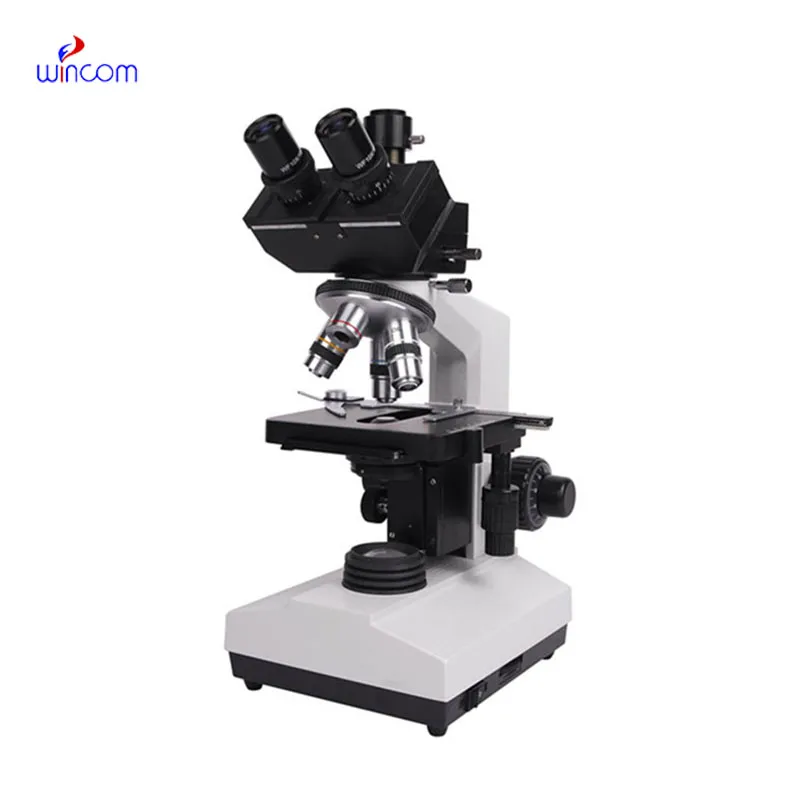
The water bath performs consistently and maintains a stable temperature even during long experiments. It’s reliable and easy to operate.
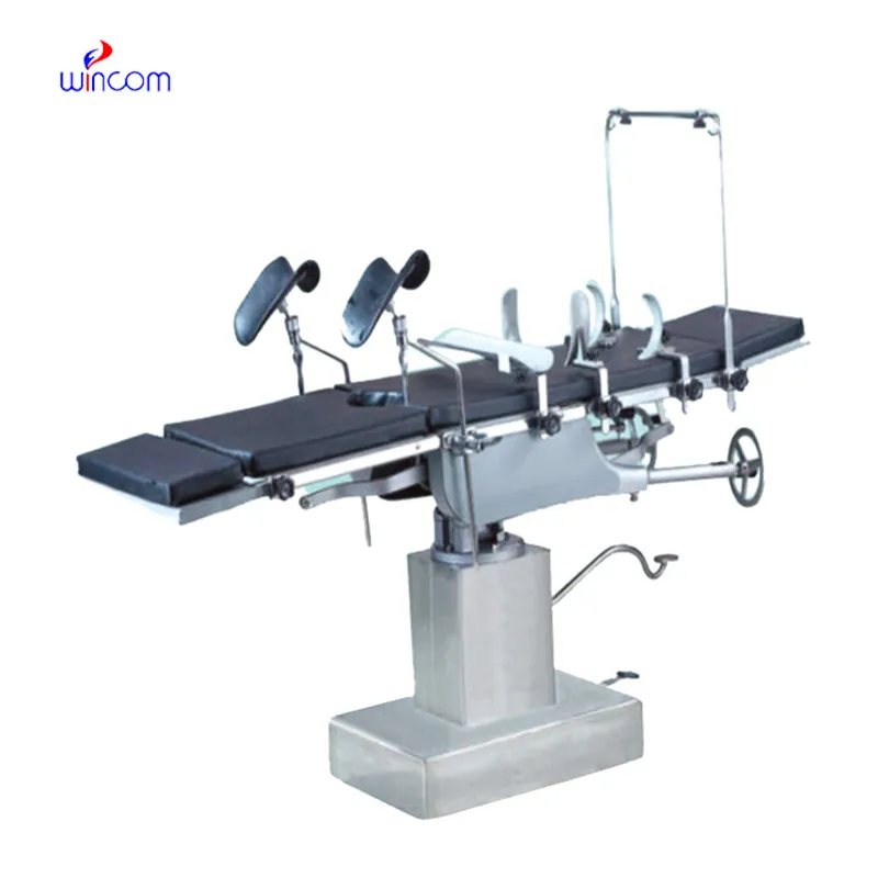
The delivery bed is well-designed and reliable. Our staff finds it simple to operate, and patients feel comfortable using it.
To protect the privacy of our buyers, only public service email domains like Gmail, Yahoo, and MSN will be displayed. Additionally, only a limited portion of the inquiry content will be shown.
We are planning to upgrade our imaging department and would like more information on your mri machin...
Could you share the specifications and price for your hospital bed models? We’re looking for adjus...
E-mail: [email protected]
Tel: +86-731-84176622
+86-731-84136655
Address: Rm.1507,Xinsancheng Plaza. No.58, Renmin Road(E),Changsha,Hunan,China

 af
af
 es
es
 ar
ar
 tr
tr
 sw
sw
 pt
pt
 th
th
 ur
ur
 bn
bn
 ne
ne
 vi
vi
 km
km
 lo
lo
 de
de
 ru
ru
 fi
fi
 nl
nl
 fa
fa
 fr
fr
 ko
ko