hplc column hangs the hospital laboratory in the sense of getting quick and reproducible results for patient sample analysis. Its use is widespread to separate small molecules, hormones, and therapeutic drugs with pinpoint accuracy. Lab staff apply hplc column in discovering biomarkers, doing pharmacokinetic studies, and metabolite profiling. Its flexibility makes it suitable for clinical applications with different requirements like research, routine diagnostics, and patient care. So, when hospitals include hplc column into their laboratory processes, they get not only the speed but also the dependable analytical performance over various departments.

hplc column allows the personnel of hospitals and laboratories to keep an eye on the presence of environmental pollutants in sterile drugs. It purifies and recognizes the remaining solvents, preservatives, and other possible impurities thus, confirming safety and meeting the requirements of regulatory authorities. This technology is vital in the battle against exposing patients to toxic agents.

hplc column is expected to have an increasing role in personalized medicine, analyzing complicated biomarkers swiftly. In the future, their application in hospitals will be centered on integrating pharmacokinetics, metabolomics, and monitoring, helping medical practitioners have access to swift and comprehensive data. The workflow in laboratories is expected to be organized.

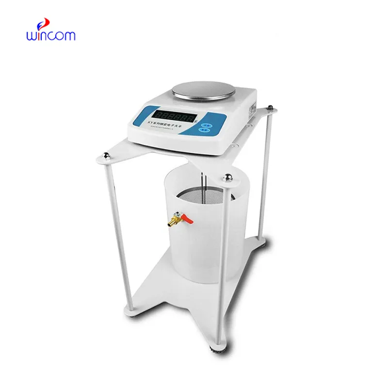
Systematic cleaning, pressure monitoring, and timely worn parts replacement are among the measures to be taken in the hospital laboratories to keep hplc column under control. Laboratory staff must ensure the observance of the suggested operating conditions, avoid the formation of air bubbles in the system, and check for proper solvent compatibility. Regular maintenance maintains the performance of the column, avoids contamination, and allows the analysis to be precise and reproducible, thereby benefiting not only routine patient testing but also experimental research.
hplc column are a major factor in the daily activities of pharmaceutical labs, as they are used for verifying drug formulations, detecting impurities, and making sure that quality standards are met. It provides accurate quantification by separating active ingredients from excipients. Lab scientists utilize this for process optimization and stability evaluation under varied conditions. By providing reproducible analytical data, hplc column assists in both method validation and research development. Its accuracy guarantees that pharmaceutical products will be compliant with regulations. In lab environments, hplc column is a time-saving method not only for compound profiling but also for comprehensive analyses, thus being a fundamental tool in the quality control of pharma and research labs dealing with drug development.
Q: What are the main parts of a microscope? A: The key components include the eyepiece, objective lenses, stage, focusing knobs, and illumination system, all working together to magnify and clarify specimens. Q: How do you clean the lenses of a microscope? A: Lenses should be cleaned using soft lens paper or microfiber cloth with a small amount of lens cleaner to avoid scratching or damaging optical coatings. Q: What magnification levels can a microscope achieve? A: Depending on the model, a microscope can typically achieve magnifications ranging from 40x to over 1000x for detailed observation of microscopic structures. Q: Why is light adjustment important in a microscope? A: Proper light adjustment ensures accurate contrast and brightness, allowing clear observation without distortion or glare during viewing. Q: Can a microscope be used for educational purposes? A: Yes, microscopes are widely used in classrooms and laboratories to teach students about biology, materials science, and microscopic analysis.
The microscope delivers incredibly sharp images and precise focusing. It’s perfect for both professional lab work and educational use.
I’ve used several microscopes before, but this one stands out for its sturdy design and smooth magnification control.
To protect the privacy of our buyers, only public service email domains like Gmail, Yahoo, and MSN will be displayed. Additionally, only a limited portion of the inquiry content will be shown.
We’re currently sourcing an ultrasound scanner for hospital use. Please send product specification...
We’re interested in your delivery bed for our maternity department. Please send detailed specifica...
E-mail: [email protected]
Tel: +86-731-84176622
+86-731-84136655
Address: Rm.1507,Xinsancheng Plaza. No.58, Renmin Road(E),Changsha,Hunan,China

 af
af
 es
es
 ar
ar
 tr
tr
 sw
sw
 pt
pt
 th
th
 ur
ur
 bn
bn
 ne
ne
 vi
vi
 km
km
 lo
lo
 de
de
 ru
ru
 fi
fi
 nl
nl
 fa
fa
 fr
fr
 ko
ko