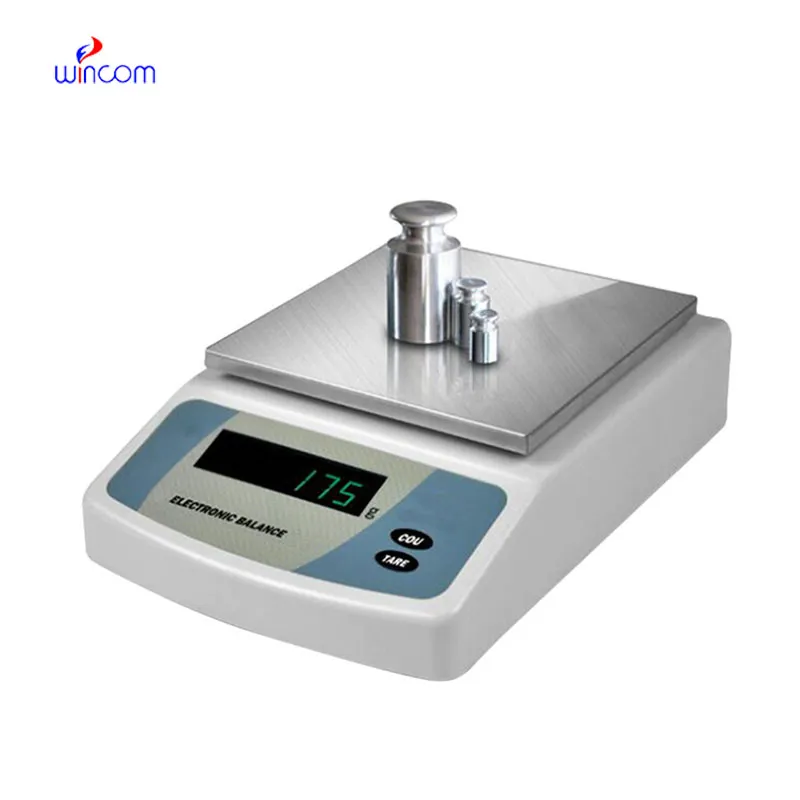
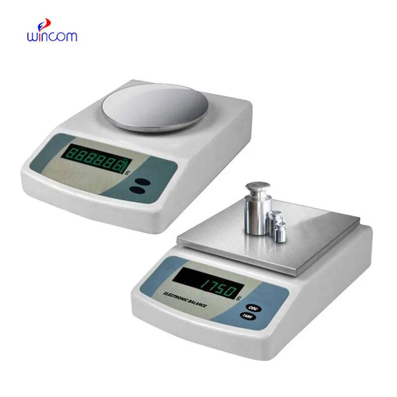
In hospital and research facility settings, sartorius analytical balance provides the critical mass measurement that is needed for delicate analyses. It is the case that reagents, samples, and medicines are weighed with the highest level of precision. Laboratory staff regard the sartorius analytical balance as their helper in making the measurements, carrying out calibrations, and performing quality assurance. Besides being of great assistance in the above activities and ensuring accurate measurement in clinical diagnosis, experimental research, and drug response monitoring, sartorius analytical balance also enhances overall laboratory performance and has a positive impact on the dependability of analytical results.

In research labs of the biomedical field, sartorius analytical balance is used while standardizing the experimental samples. For the purpose of testing, h researchers have to measure the biological or chemical samples very accurately and in this way, they do not use more than the required amount of sample for analytical testing. This process keeps the studies that compare different methodologies consistent and at the same time it prevents different results that are due to the difference in the samples’ mass. By providing correct input values, sartorius analytical balance makes it easier for the experimenters to repeat the experiments and to trust the data more in the hospitals’ research institutions.
The future usage of sartorius analytical balance in hospitals will mainly be geared towards the compatibility of automation. It is predicted that analytical balances will connect with the robotic sample handling mechanisms of clinical and pharmaceutical labs. This connection will allow for uninterrupted work, lesser operator involvement, and better consistency. The scale of La due to the hospitals aiming for total automation, sartorius analytical balance will be your basic measuring unit in these cutting-edge platforms.
The maintenance of sartorius analytical balance involves the aspects of storage and inactivity care that come first. The balance should be protected from dust and vibration when it is not in active use. Periodically checking the operational status during long storage prevents unnoticed performance drift. These practices guarantee that sartorius analytical balance is still capable of accurate use in laboratories, medical and hospital settings.
sartorius analytical balance is employed in hospital labs for the reliable quality control of reagents, chemicals, and medications. Its exactness provides accurate concentrations for assays, patient treatments, and experimental protocols. The laboratory personnel regularly calibrate sartorius analytical balance to rule out mistakes. Its application keeps the standard of hospital laboratories, allows the reproducibility, and builds trust in clinical and research outcomes.
Q: What maintenance does an Analytical Balance require? A: A periodic cleaning, checking of the calibration, and also verifying the performance are all necessary. Q: Can an Analytical Balance handle continuous daily use? A: Yes, provided that the correct laboratory conditions and rules are followed. Q: Why is leveling important for an Analytical Balance? A: The accuracy and repeatability of the measurements depend on proper leveling. Q: Can Analytical Balances be connected to laboratory systems? A: Most of the models allow connectivity with laboratory information systems. Q: Are Analytical Balances sensitive to vibration? A: Yes, stable weight readings can be disturbed by vibrations.
The microscope delivers incredibly sharp images and precise focusing. It’s perfect for both professional lab work and educational use.
I’ve used several microscopes before, but this one stands out for its sturdy design and smooth magnification control.
To protect the privacy of our buyers, only public service email domains like Gmail, Yahoo, and MSN will be displayed. Additionally, only a limited portion of the inquiry content will be shown.
Hello, I’m interested in your water bath for laboratory applications. Can you confirm the temperat...
I’d like to inquire about your x-ray machine models. Could you provide the technical datasheet, wa...
E-mail: [email protected]
Tel: +86-731-84176622
+86-731-84136655
Address: Rm.1507,Xinsancheng Plaza. No.58, Renmin Road(E),Changsha,Hunan,China

 af
af
 es
es
 ar
ar
 tr
tr
 sw
sw
 pt
pt
 th
th
 ur
ur
 bn
bn
 ne
ne
 vi
vi
 km
km
 lo
lo
 de
de
 ru
ru
 fi
fi
 nl
nl
 fa
fa
 fr
fr
 ko
ko