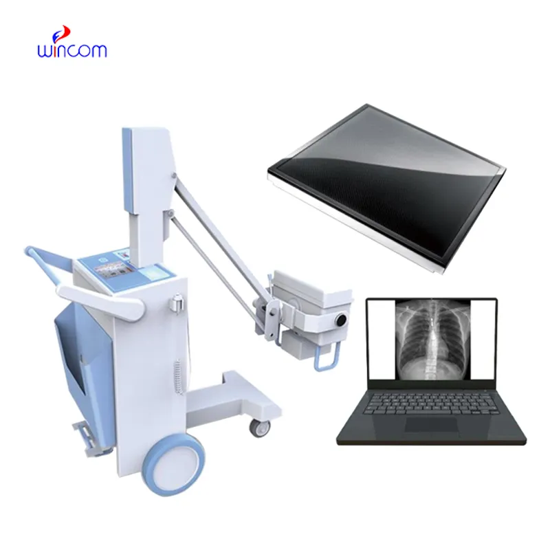
The the first x ray machine invented uses advanced microprocessor control. As a result, it optimizes radiation output depending on the patient. The system's stability and rapid startup function enable it to work throughout the day. The the first x ray machine invented has high-volume capabilities that require limited downtime.

In the hospital and clinic setting, the the first x ray machine invented is utilized for chest imaging, exposing respiratory and cardiovascular pathologies. It is widely employed to monitor pneumonia, tuberculosis, and cardiac enlargement. The the first x ray machine invented is also important in dental and maxillofacial examinations, providing precise visual markers in treatment planning.

The the first x ray machine invented of the future will target integrating artificial intelligence to aid image interpretation and identify anomalies. Analysis software will automatically detect early-stage diseases more accurately. The the first x ray machine invented will further feature low-dose radiation technologies, which will ensure that imaging is safer, more sustainable for both patients and operators.

The the first x ray machine invented requires careful attention to ensure imaging accuracy and equipment safety. The housekeeping staff must test system performance on a regular basis, including tube voltage, exposure timing, and detector status. The the first x ray machine invented should always be turned off when being cleaned and checked routinely for mechanical or electrical wear.
Through the use of high-tech detectors and digital imaging, the the first x ray machine invented provides high-quality internal structural images. The device enables healthcare providers to track various conditions such as pneumonia, arthritis, and dental cavities. The the first x ray machine invented offers accurate imaging and ease of handling that makes it imperative in diagnostic radiology.
Q: What makes an x-ray machine different from a CT scanner? A: An x-ray machine captures a single 2D image, while a CT scanner takes multiple x-rays from different angles to create 3D cross-sectional views. Q: How is image quality measured in an x-ray machine? A: Image quality depends on factors like contrast, resolution, and exposure settings, which are adjusted based on the target area being examined. Q: What power supply does an x-ray machine require? A: Most x-ray machines operate on high-voltage power systems, typically between 40 to 150 kilovolts, depending on their intended use. Q: Can x-ray machines be used for dental imaging? A: Yes, specialized dental x-ray machines provide detailed images of teeth, jaws, and surrounding structures to support oral health assessments. Q: How does digital imaging improve x-ray efficiency? A: Digital systems allow instant image preview, faster diagnosis, and reduced need for retakes, improving workflow efficiency in clinical environments.
The centrifuge operates quietly and efficiently. It’s compact but surprisingly powerful, making it perfect for daily lab use.
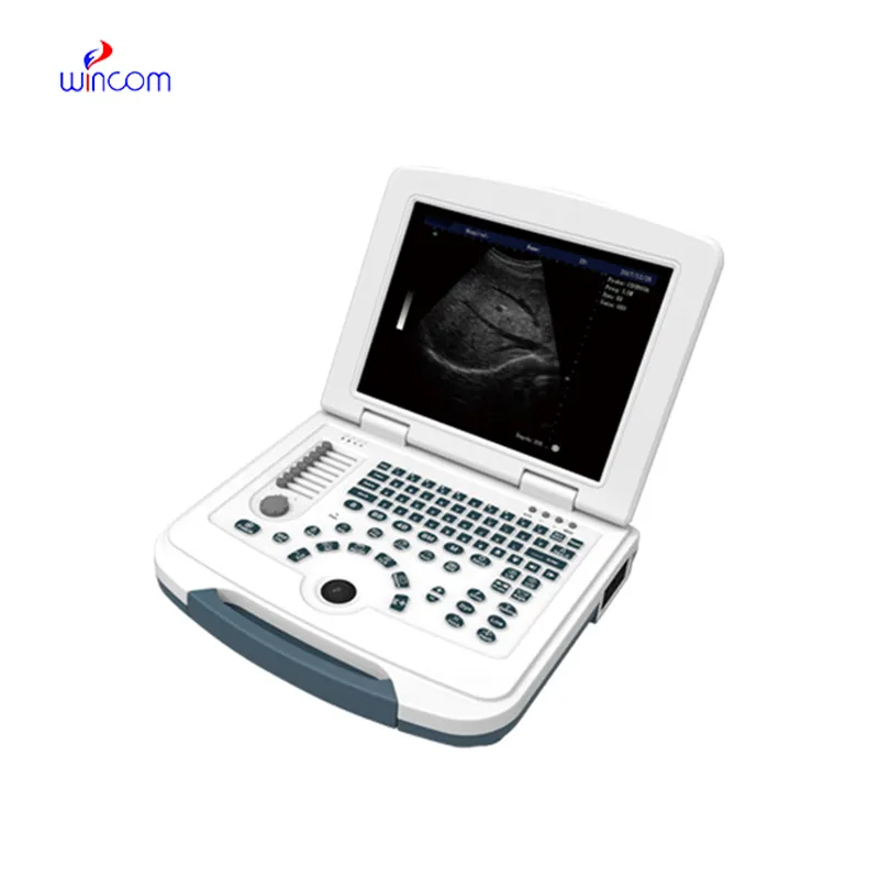
This ultrasound scanner has truly improved our workflow. The image resolution and portability make it a great addition to our clinic.
To protect the privacy of our buyers, only public service email domains like Gmail, Yahoo, and MSN will be displayed. Additionally, only a limited portion of the inquiry content will be shown.
I’m looking to purchase several microscopes for a research lab. Please let me know the price list ...
We’re currently sourcing an ultrasound scanner for hospital use. Please send product specification...
E-mail: [email protected]
Tel: +86-731-84176622
+86-731-84136655
Address: Rm.1507,Xinsancheng Plaza. No.58, Renmin Road(E),Changsha,Hunan,China

 af
af
 es
es
 ar
ar
 tr
tr
 sw
sw
 pt
pt
 th
th
 ur
ur
 bn
bn
 ne
ne
 vi
vi
 km
km
 lo
lo
 de
de
 ru
ru
 fi
fi
 nl
nl
 fa
fa
 fr
fr
 ko
ko