The virus under microscope is engineered to deliver consistent performance at all magnification levels. With precision focusing knobs and a rugged mechanical stage, it offers accurate sample positioning and smooth handling. The illumination system provides even lighting for clear observation of opaque and transparent specimens. Most virus under microscope models have modular configurations, which can be customized for particular fields like biology, metallurgy, or semiconductor inspection.

Across the worlds of science, industry, and education, the virus under microscope enables research at the microscopic level. It is an essential tool in medical diagnosis to analyze blood, tissues, and pathogens. Environmental scientists apply the virus under microscope to determine bacteria and microalgae that indicate water levels of quality. In materials science, it enables nanostructure analysis and the identification of defects. Art conservators apply the virus under microscope to analyze pigments and varnish layers. Its ability to produce accurate, detailed imagery makes it a valuable resource in continuing discovery and research development.

With the progress of technology, the virus under microscope will turn into a smarter and more interactive research tool. Compatibility with AI will allow it to detect patterns, recognize anomalies, and measure data automatically. The virus under microscope will also make remote diagnostics possible, where the samples from every corner of the world can be diagnosed remotely by specialists. Advances in imaging sensors and optical systems will provide better depth resolution and faster capture rates. These will expand the uses of the virus under microscope in medicine, nanotechnology, and education.

Users should implement a routine maintenance plan to ensure the virus under microscope remain in excellent working condition. Clean all optical parts using a blower or soft brush initially before a thorough cleaning. Do not disassemble the instrument at any time save by qualified individuals. Use light lubricant on moving parts to prevent stiffness and wear. The virus under microscope should be kept in a chemical fume and moisture-free environment. Power cables and lighting systems should be checked regularly for signs of premature deterioration or breakdown.
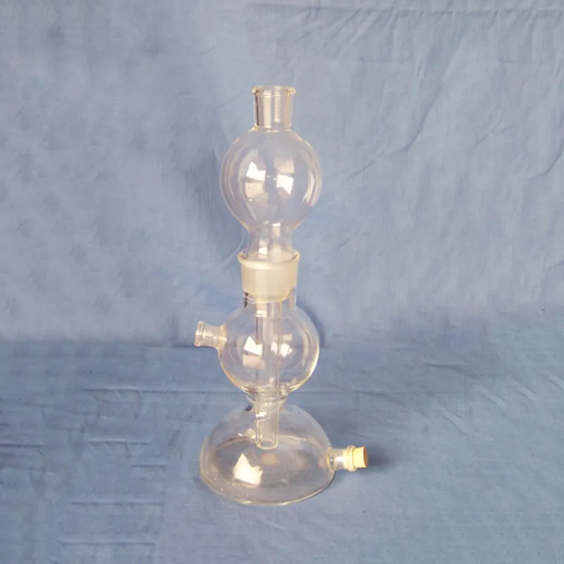
With a virus under microscope, human man can explore the microcosm with unprecedented clarity. The instrument magnifies small samples so that exact study can be conducted in laboratories, clinics, and schools. The virus under microscope recognizes cell morphology, bacterial cultures, and intricate material surfaces. Although optical and electronic technology has been enhanced, the virus under microscope of today's time offers more magnification, image stability, and integration into digital media for efficient data registration and perception.
Q: How do environmental conditions affect a microscope? A: Excessive heat, moisture, or dust can damage optical and mechanical components, so the microscope should be used in a clean, controlled environment. Q: Can a microscope capture images or videos? A: Many modern microscope models include digital cameras that enable high-resolution image and video capture for documentation or analysis. Q: What training is required to operate a microscope? A: Basic understanding of optics and focusing principles is recommended, though most educational microscopes are designed for simple, intuitive use. Q: Why is regular maintenance important for a microscope? A: Regular maintenance prevents dust buildup, mechanical wear, and misalignment, ensuring consistent performance and image clarity. Q: Can a microscope be used outside the laboratory? A: Portable and handheld microscope models are available for field studies, allowing researchers to observe and analyze samples on site.
This ultrasound scanner has truly improved our workflow. The image resolution and portability make it a great addition to our clinic.
The delivery bed is well-designed and reliable. Our staff finds it simple to operate, and patients feel comfortable using it.
To protect the privacy of our buyers, only public service email domains like Gmail, Yahoo, and MSN will be displayed. Additionally, only a limited portion of the inquiry content will be shown.
Could you share the specifications and price for your hospital bed models? We’re looking for adjus...
We are planning to upgrade our imaging department and would like more information on your mri machin...
E-mail: [email protected]
Tel: +86-731-84176622
+86-731-84136655
Address: Rm.1507,Xinsancheng Plaza. No.58, Renmin Road(E),Changsha,Hunan,China

 af
af
 es
es
 ar
ar
 tr
tr
 sw
sw
 pt
pt
 th
th
 ur
ur
 bn
bn
 ne
ne
 vi
vi
 km
km
 lo
lo
 de
de
 ru
ru
 fi
fi
 nl
nl
 fa
fa
 fr
fr
 ko
ko