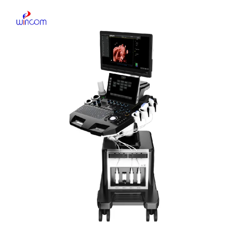
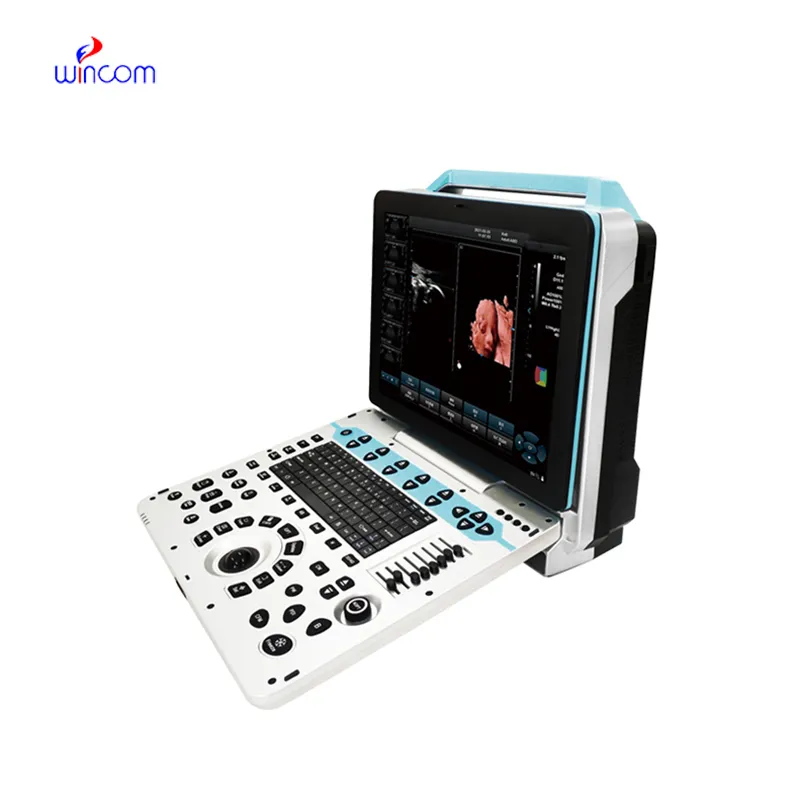
The wireless ultrasound scanner, being designed for flexibility, brings the user a variety of imaging modes such as B-mode, M-mode, and color Doppler to use. Because of its small size, it can be easily moved from one place to another and that is why it can be used for examining patients in bed. The wireless ultrasound scanner provides good imaging quality, which can be depended upon in cases of routine diagnostics, fieldwork, and emergency medical interventions.

The vast clinical applications of the wireless ultrasound scanner technology made it possible for nephrology to monitor kidney function efficiently and detect abnormalities in kidney structure. In the frontiers of endocrinology, the obtained data can reveal even the smallest nodules in the glands. The wireless ultrasound scanner is also a surgical device for blood flow patterns and vessel integrity.

The wireless ultrasound scanner should integrate with intelligent diagnostic ecosystems and communicate effortlessly with smartphones and electronic records. The synchronized exchange of data in real-time should enable constant patient observation. The next version should focus on improved design, better processing power of artificial intelligence algorithms, and enhanced reconstruction functions.

The wireless ultrasound scanner require a certain set of procedures when it comes to handling them. The images on the display should always be cleaned with soft, lint-free cloths. The probe membranes should always be checked for the absence of cracks. The wireless ultrasound scanner also require calibration verification.
The wireless ultrasound scanner is an essential diagnostic modality in the modern healthcare system, permitting the non-invasive imaging of internal organs and tissues. By transmitting sound waves and reading their echoes, it provides real-time data on physiology. The wireless ultrasound scanner makes precise diagnoses feasible in all specialties, improving clinical decision-making and patient confidence.
Q: What imaging modes are available on the ultrasound scannert? A: It supports multiple modes such as B-mode, M-mode, and color Doppler for diverse diagnostic applications. Q: How does the ultrasound scannert improve diagnostic accuracy? A: By providing high-resolution images and real-time feedback, it enables more precise medical evaluations. Q: Can the ultrasound scannert be used in field or remote settings? A: Yes, its portable versions are designed for mobility and can be used in clinics, hospitals, or mobile healthcare units. Q: What kind of display does the ultrasound scannert use? A: It typically features a high-definition digital display that enhances image visualization and readability. Q: How is data from the ultrasound scannert managed? A: The device allows secure storage, easy access, and export of imaging data through USB or network connections.
The hospital bed is well-designed and very practical. Patients find it comfortable, and nurses appreciate how simple it is to operate.
This x-ray machine is reliable and easy to operate. Our technicians appreciate how quickly it processes scans, saving valuable time during busy patient hours.
To protect the privacy of our buyers, only public service email domains like Gmail, Yahoo, and MSN will be displayed. Additionally, only a limited portion of the inquiry content will be shown.
Hello, I’m interested in your water bath for laboratory applications. Can you confirm the temperat...
I’m looking to purchase several microscopes for a research lab. Please let me know the price list ...
E-mail: [email protected]
Tel: +86-731-84176622
+86-731-84136655
Address: Rm.1507,Xinsancheng Plaza. No.58, Renmin Road(E),Changsha,Hunan,China

 af
af
 es
es
 ar
ar
 tr
tr
 sw
sw
 pt
pt
 th
th
 ur
ur
 bn
bn
 ne
ne
 vi
vi
 km
km
 lo
lo
 de
de
 ru
ru
 fi
fi
 nl
nl
 fa
fa
 fr
fr
 ko
ko